club foot horse x ray
The equine club foot is defined as a hoof angle greater than 60 degrees. Proceedings 32nd Annual Meeting Am Assoc Equine Pract 1986 32.
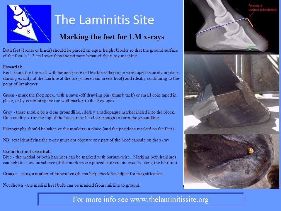
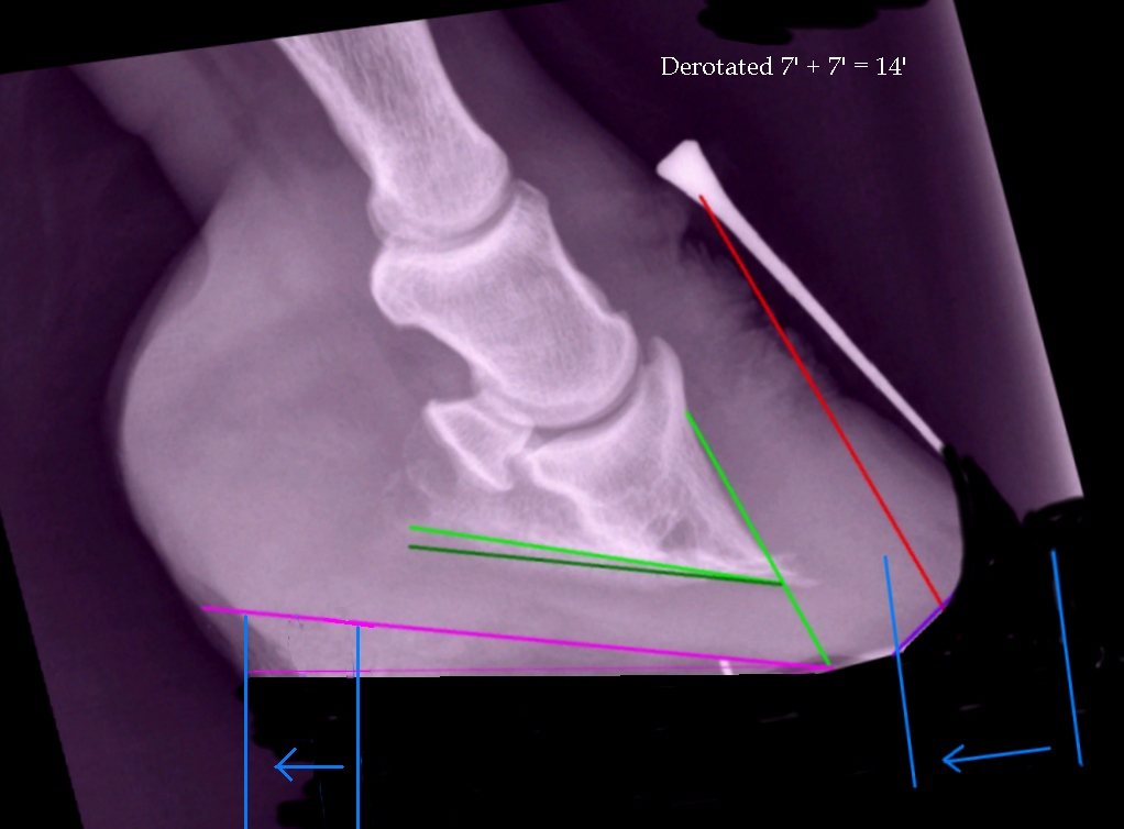
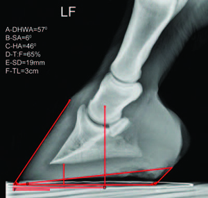
Understanding X Rays The Laminitis Site
MedWell Testosterone Weight Loss Hormone Replacement 201 806-6099.
. As the foot grows out in these horses there is a propensity for the dorsal wall to distort and flare. Home Practice Team Contact 201 806-6099. For example lets say you have a grade 2 club foot.
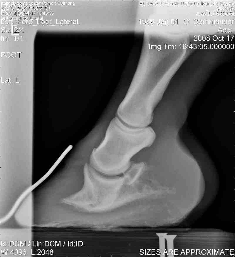
Radiographs of the equine foot or feet are a standard diagnostic used for hoof or foot related problems and in lameness exams because a high percentage of lameness originates in. MECHANICAL LAMINITIS TREATMENT. Lateral talocalcaneal angle.
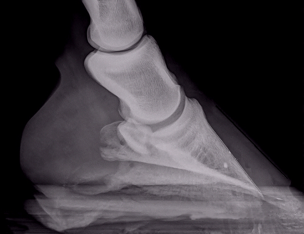
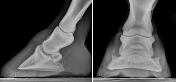
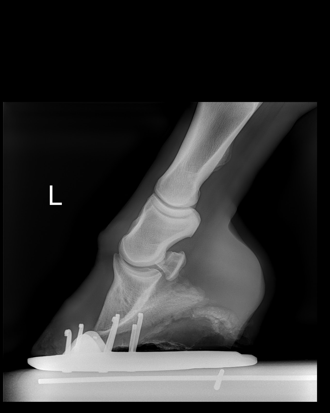
In a club foot the angle of the hoof and pastern in relation to the ground is abnormally steep. There are varying degrees of club foot. Talocalcaneal parallelism is the radiographic feature of clubfoot talipes.
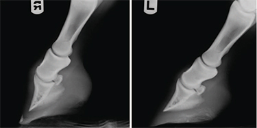
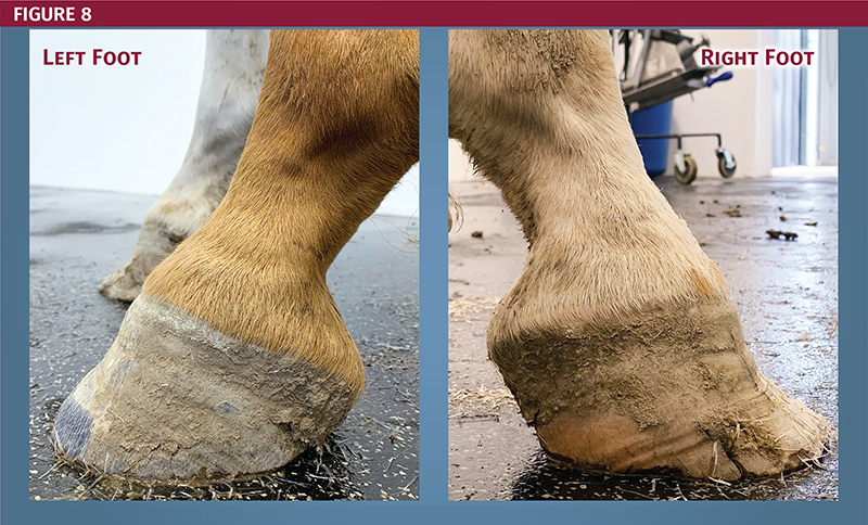
Podiatry in equine veterinary practice is gaining increasing attention. This horse has very healthy sound feet despite a grade 1 right front club. What we see externally as the equine clubbed foot is actually caused by a flexural deformity of the distal.
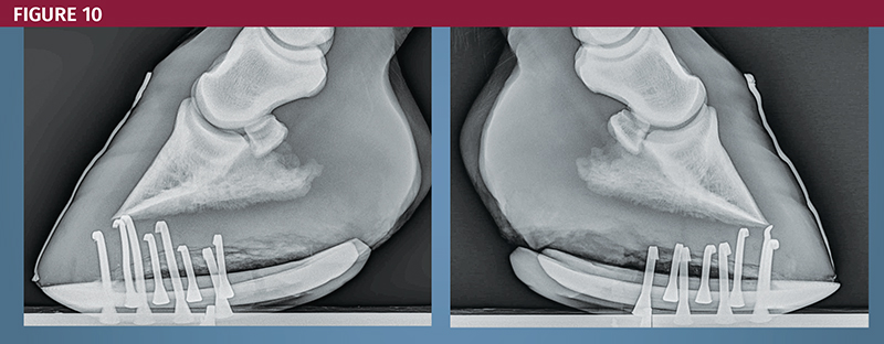
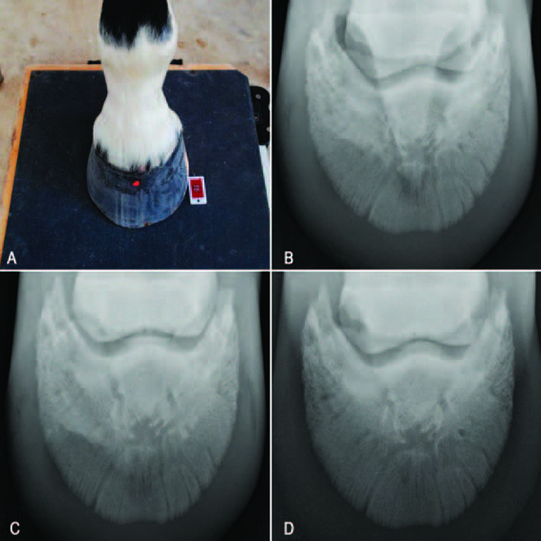
Horses feet are placed on x ray transparent lifts or blocks to enable x rays to show complete hoof including sole. The longitudinal arch is abnormally high. An X ray of your horses foot can help you predict the future while it shows you the present.
The right front growth rings. We continue to learn more about the function and biomechanics of the horses foot and develop new and. X-Ray providers in Hackensack NJ.
An X ray of your horses foot can help you predict the future while it shows you the present. Many people call the ahead using services as well as wait in the lobby for your X-Ray. The point here is to educate everyone that were not looking for a nice.
Radiographic changes in the navicular bone of normal horses. 225 Millburn Ave Ste 104a Millburn NJ 07041. Composition main components parts model no.
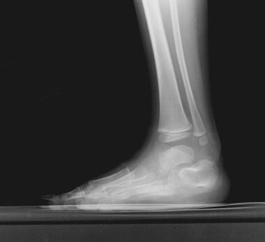
Horses feet are placed on x ray transparent lifts or blocks to enable x rays to show complete hoof including sole. One of the most common of all birth defects. Occasionally radiographs are necessary to diagnose clubfeet associated with tibial hemimelias.
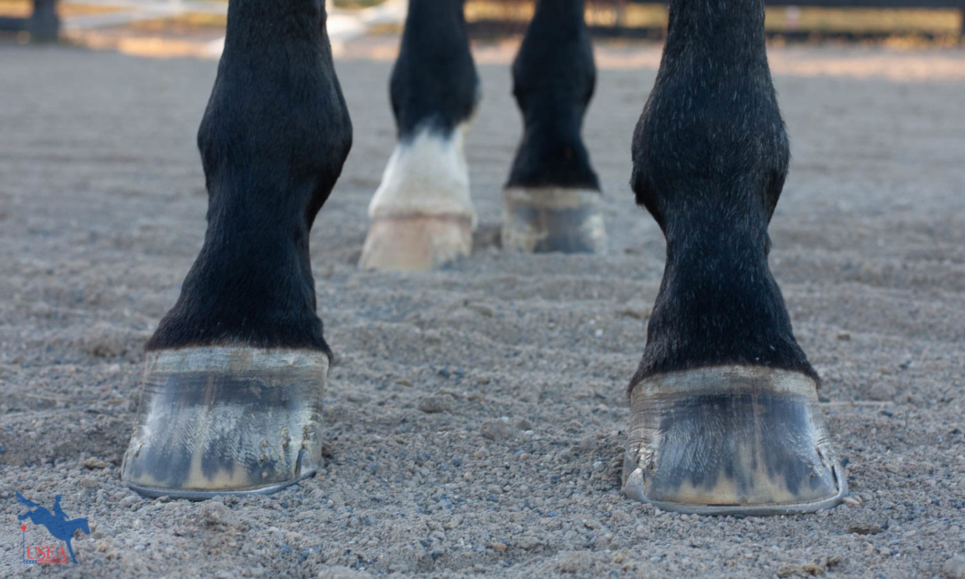
A club foot horse is typically recognized and defined as having one front hoof growing at a much steeper angle than the other with a short dished toe very high heels. Ray Paulick 859 312-2102. But what does the term mean and what actually constitutes a clubfoot on a horse.
Generally the greater the upright angle the more severe the club foot. The right front growth rings indicate a tendency for a negative PA in the. Turner TA Kneller SK Badertscher RR et al.
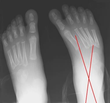
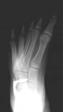
The horse should be stood on a flat level surface. X-ray of feet typical clubfoot Clubfoot Introduction. Emily Alberti 859 913-9633.
Club Foot Heritability in Horses. The normal range of hoof angle is 50 to 55 degrees while a club. It is more available from the need to X-ray for any type of injury with the get checked out.
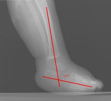
Lateral radiograph of the right foot shows that the long axes of the talus and calcaneus are nearly parallel. A matter of degree. To appreciate bone position the radiographs should be taken with the horse bearing weight and both feet placed on wooden blocks of equal.
Comes very useful in horses with upright feet the best example being the club-footed horse. In the past the condition was defined as any hoof angle that. Figure 1A Left.
Clubfoot talipes equinovarus TEV is one of the major orthopedic conditions of childhood. Talipes equinovarus consists of four elements 7.
Staying Sound The Importance Of Foot Balance
High Heels The Laminitis Site

Natural Angle Volume 15 Issue 1 Spanish Lake Blacksmith
Thrush Cured Cowboy Pads And Copper Sulfate
Equine Podiatry Dr Stephen O Grady Veterinarians Farriers Books Articles

How To Treat Club Feet And Closely Related Deep Flexor Contraction
Understanding X Rays The Laminitis Site
Clubfoot Imaging Practice Essentials Radiography Computed Tomography
Clubfoot Imaging Practice Essentials Radiography Computed Tomography
Equine Therapeutic Farriery Dr Stephen O Grady Veterinarians Farriers Books Articles
Clubfoot Imaging Practice Essentials Radiography Computed Tomography
Clubfoot Imaging Practice Essentials Radiography Computed Tomography

Being Able To Read X Rays Of The Equine Documentalist Facebook
Recognizing And Managing The Club Foot In Horses Horse Journals
Equine Therapeutic Farriery Dr Stephen O Grady Veterinarians Farriers Books Articles
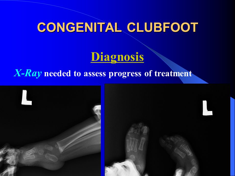
Congenital Clubfoot Congenital Talipes Equino Varus Ppt Video Online Download
Low Foot Case Study Dixie S Farrier Service

Equine Podiatry In Wendell Nc Neuse River Equine Hospital
Equine Therapeutic Farriery Dr Stephen O Grady Veterinarians Farriers Books Articles